

- Name
- Hannah Robbins
- hlrobbins@jhu.edu
- Cell phone
- 667-232-9047
Cells and tissues surrounding a breast cancer tumor may hold critical information about how patients will respond to treatment, according to a new study from Johns Hopkins University.
The research represents a significant step toward developing artificial intelligence tools that could help oncologists more accurately determine prognoses and decide which treatments may be most effective on a case-by-case basis.
Key Takeaways
- Cancerous and noncancerous cells surrounding a tumor make up its microenvironment.
- Researchers developed an AI tool that could analyze hundreds of breast cancer tumor microenvironments and detect recurring patterns.
- Patients with similar tumor microenvironments often had similar prognostic outcomes.
"When we can identify recurring elements in the microenvironments—cancerous cells, specific types of noncancerous cells, and their patterns of spatial organization—they're like little flags saying, 'Turn your attention over here, we may be important,'" said senior author Jeremias Sulam, an assistant professor of biomedical engineering who studies machine learning at Johns Hopkins University. "Our study is about identifying patterns and biomarkers that could help us understand how the disease progresses."
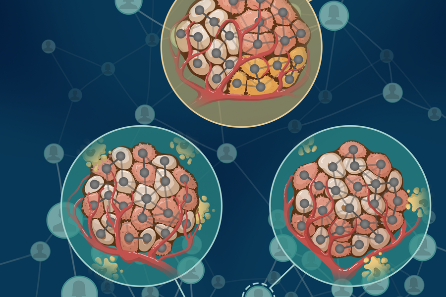
The research was featured on the cover of the March issue of Patterns. While the researchers stressed that the technology is still some time away from directly enhancing patient care, they said it holds great promise for improving breast cancer treatments.
To gain a better understanding of tumor environments, the researchers created an interpretable machine learning model that analyzed images of microscopic tissues and biopsy samples from 579 breast cancer patients undergoing standard treatments such as chemotherapy. The model looked for patterns in the tissue within and surrounding the tumors, noting every cell's position in relation to the tumors and their interactions with other neighboring cells.
The algorithm found 66 distinct cell patterns in the tumor microenvironments and clustered patients with similar patterns into seven groups, some of which ended up having better prognostic outcomes than others.
Patients in the group with the best survival outcomes had one thing in common: a mixture of three different cells (CK8-18high, CXCL12high, and CK+CXCL12+). Meanwhile, patients with self-aggregated HER2+ tumor cells had the worst treatment outcomes—unsurprising but reassuring, the researchers said, because the HER2 gene has been associated with aggressive breast cancers and low survival rates.
Triple negative breast cancer patients who had a similar pattern of well-organized immune cells surrounding their tumors had better survival rates than others with the same disease. The findings suggest there may be crucial information in those patterns that oncologists could use for prognoses, the researchers said.
Because the researchers developed an interpretable machine learning model, they were able to determine the specific elements that made up the patterns in the microenvironments.
Most modern algorithms are like black boxes—they can group or classify data, but they're hard to interpret and can't tell you which parts of the data are driving the algorithm's decisions, the researchers said. It's like throwing a bunch of soup ingredients into a pot and not knowing which are most important to the flavor. This new methodology allowed the researchers to go back to the results and pinpoint exactly which characteristics are influencing the results.
"We're developing a toolbox that you can use anytime you have a massive data set and you don't want to start with an explicit hypothesis," said first author Zhenzhen Wang, a PhD candidate in biomedical engineering at Johns Hopkins University. "Typically, you'd have to design a study to ask if one specific pattern of cells is important, and check if the answer is yes or no. But with our model, we can save time by asking open-ended questions about which patterns are important when trying to understand how patients will respond to treatment or have a better [chance of] survival."
Sulam added: "Once we identify the patterns, oncologists and wet lab scientists can follow up to see what mechanisms might underlay these patterns, which could in turn provide directions as to how to intervene with a more targeted treatment."
In the future, the research group plans to use this methodology for other types of imaging technologies and to find drivers of other types of cancer.
Other authors include Cesar Santa-Maria, an associate professor of oncology, and Aleksander Popel, a professor of biomedical engineering and oncology, both from the Johns Hopkins University School of Medicine.
The work was supported by funding from the National Institutes of Health grant R01CA138264 and NSF CAREER Award CCF 2239787.
Posted in Science+Technology
Tagged cancer, breast cancer, biomedical engineering, artificial intelligence