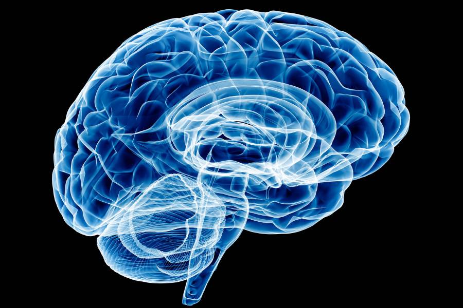
Doctors performing surgery to remove brain tumors must take care to avoid the brain's eloquent cortex. Damage to that sensitive area, located in the left temporal and frontal lobe, can be devastating, resulting in impairment of a person's ability to understand language and, sometimes, even paralysis.
Currently, the only way for surgeons to map the eloquent cortex before performing a procedure is to insert electrodes directly into the patient's brain, a practice that is both highly traumatic and invasive.
A doctoral candidate at Johns Hopkins University is developing an alternative approach that uses painless, non-invasive functional magnetic resonance imaging, or fMRI, to locate the eloquent cortex pre-surgery. The fMRI scanning technique creates a video of the brain through measurement of blood flow over time.

Image caption: Naresh Nandakumar
"Our method improves upon the use of electrodes by working on resting-state fMRI data, which can be taken days or weeks before the surgery," said Naresh Nandakumar, a doctoral candidate in the Whiting School of Engineering's Department of Electrical and Computer Engineering and a member of the Neural Systems Analysis Laboratory. "And because this approach is non-invasive, it makes preoperative mapping techniques available to people who could not have tolerated the use of electrodes, such as young children, patients with aphasia, or patients with disabilities."
Nandakumar's model leverages the fact that regions in the brain that have synchronous blood flow patterns are more likely to be involved in similar cognitive processes. More specifically, his model applies deep learning techniques on a graph that summarizes cognitive activity connecting various regions of the brain. Deep learning is a type of artificial intelligence learning that uses neural networks modeled on the human brain to analyze hefty amounts of data to perform certain tasks or functions.
Nandakumar's model then identifies how the eloquent cortex is connected with the rest of the brain from a functional point of view, showing the surgeon which area to avoid during the procedure.
"Naresh's work focuses on resting-state fMRI, which is a passive and easily acquired imaging modality," said Archana Venkataraman, assistant professor in the Department of Electrical and Computer Engineering and also the principal investigator for the NSA Laboratory. "He has developed a robust deep learning framework that leverages network interactions within this data for reliable language and motor mapping, thus helping clinicians to plan safer and more effective neurosurgeries."
Nandakumar believes that adding more data to his model will enhance its ability to accurately map the eloquent cortex. He plans to use data from diffusion tensor imaging, which identifies the white matter fiber tracts, or "highways" in the brain through which blood flows, to further examine the brain's structural connectivity profile and create a connectivity graph, improving his model's results.
Nandakumar comes by his interest in neuroimaging naturally; both his father and sister are neurologists.
"Tackling a relatively unsolved problem with novel machine and deep learning methods is very exciting to me," he said, noting that Johns Hopkins has proven to be "the perfect place" to pursue those interests in a truly cross-disciplinary way.
"The collaboration my lab has with the Neuroradiology Division at the JHU School of Medicine is what made this work possible," Nandakumar said. "This dataset is very specialized, and the experts that we work with have a lot of insight into the biological aspect of our work, which has been tremendously helpful."
Posted in Science+Technology
Tagged electrical and computer engineering, fmri, brain surgery, neuroimaging


