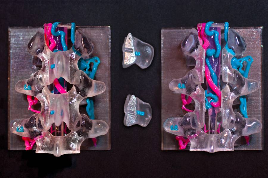
This educational model of a spinal perimedullary arteriovenous fistula is a 3D print showing the precise location of a fistula by combining MRI data of the spinal cord with 3D angiography data of the bone and vasculature. Created by Lydia Gregg, associate professor in the School of Medicine and certified medical illustrator, the model allows interventional neuroradiologists, neurosurgeons, and caregivers to show patients how fistulas involve direct connections between arteries (in magenta) and veins (in blue) at the surface of the spinal cord.
Posted in Science+Technology
Tagged 3-d printing


