- Name
- Roberto Molar Candanosa
- rmolarc1@jh.edu
- Office phone
- 443-997-0258
- Cell phone
- 443-938-1944
By genetically testing nearly 1,000 embryos, scientists have provided the most-detailed analysis of embryo fate following human in vitro fertilization.
Nearly half the embryos studied underwent developmental arrest because of genetic mishaps in early development—a revealing insight that suggests more IVF babies could come to term with changes in the fertility treatment process. The unique combination of data from arrested embryos also sheds new light on the still largely mysterious earliest stages of pregnancy through natural means.

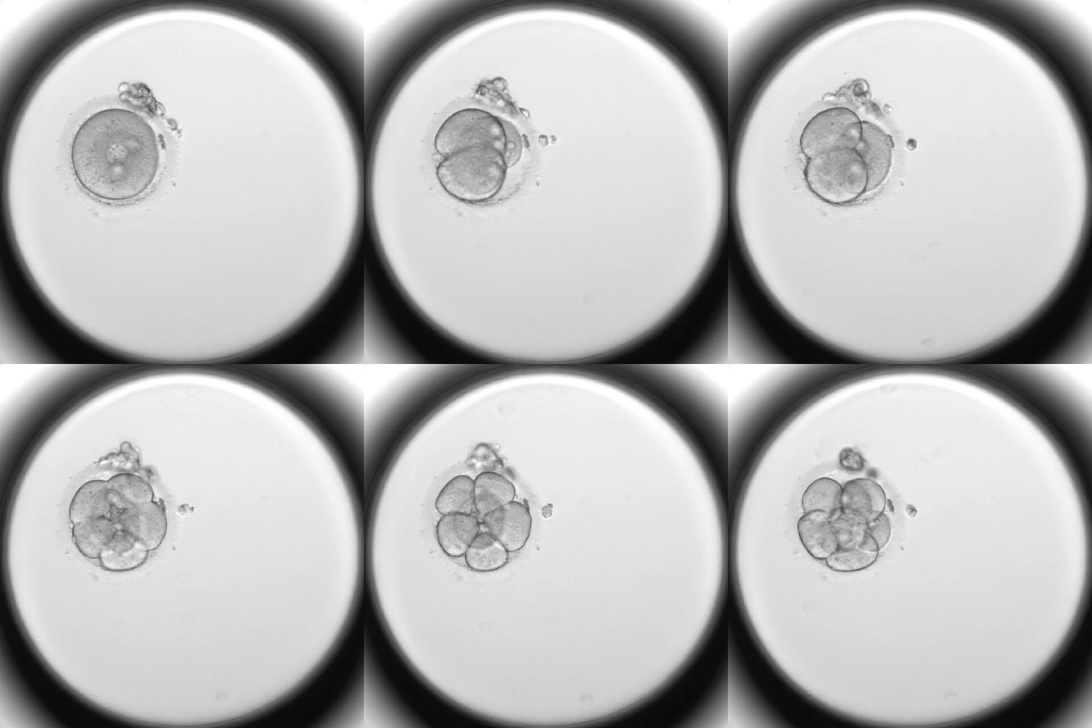
Image caption: A time lapse clip of a common type of abnormal cell division, where an embryo cleaves directly from a single-celled zygote into three (rather than two) cells. The new research shows such abnormal division is strongly associated with chromosome abnormalities and embryo arrest.
Image credit: Christian Ottolini.
"We think this also happens in natural conception, and it's why it takes, on average, several or more months to get pregnant," said author Rajiv McCoy, an assistant professor of biology at Johns Hopkins University. "It is very surprising that most of these embryo arrests are coming not from errors in egg formation but from errors happening in cell divisions after fertilization. The fact that these errors don't come from the egg suggests that maybe they could be mitigated by changing the way IVF is done."
The research was published today in Genome Medicine.
Looking for genetic differences, Johns Hopkins and London Women's Clinic researchers in the UK compared IVF embryos that failed to develop within a few days of fertilization with embryos that survived.
"Genetic testing is typically only done on IVF embryos that survive in order to decide which embryo to transfer to the uterus," McCoy said. "But from a biology standpoint, if we want to understand what's allowing these embryos to survive, then we have to test all other embryos, too."
The findings reveal how some embryos start growing properly while the maternal genetic material pre-loaded into the egg control cell division, only to falter and stall when the embryo's genes take over.
Human cells typically receive 46 chromosomes, 23 from each parent. The team discovered nonviable embryos started with the 46-chromosome set but then passed down incorrect numbers of chromosomes as cells divided.
"It doesn't really matter if you have extra missing chromosomes in the very beginning because the maternal machinery is controlling things," McCoy said. "When the embryo's genome turns on, that's when things go wrong."
Human embryos experience unusually high rates of chromosome gain and loss, known as aneuploidy, in early development. Scientists have studied aneuploidy for decades by screening IVF embryos, and these mishaps are well understood to be the cause of pregnancy loss in humans. Because aneuploidy is rare in many other species, McCoy said, the findings could help explain why pregnancy loss and miscarriage are so common in humans.
"Aneuploidy is an example of an extremely strong type of natural selection that's going on every generation in humans," McCoy said. "It might just be a feature of human reproduction and development, but it has implications for IVF. In the long term, we hope that we can improve genetic testing and improve IVF outcomes."
The researchers plan to run additional tests on specific cells from arrested embryos to trace the chromosomes' origins and see whether abnormal cell divisions are linked to maternal or paternal genetics. They also want to better understand whether factors such as the chemical composition in the dish where the embryos are grown could improve chances for survival.
"We could potentially correct a lot of these things by understanding more about the machinery that causes embryo arrest," said co-author Michael Summers, a consultant in reproductive Medicine at London Women's Clinic. "The problem could be that the chemical composition of the culture medium that is commonly used will not allow all embryos to grow, that the abnormal cell divisions are due to stresses on the egg and early embryo that causes the abnormal divisions associated with chromosome abnormalities."
Other study authors include Abeo McCollin, Kamal Ahuja, and Alan H. Handyside, of London Women's Clinic and University of Kent, and Christian S. Ottolini, of The Evewell and University College London.
This research was supported by the National Institute of General Medical Sciences of the National Institutes of Health under Award R35GM133747.
Posted in Science+Technology


