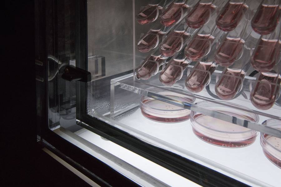
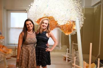
At the Empty Gallery, an art space in Hong Kong, a clear plastic cube illuminated by bright lights housed glass tubes filled with mysterious liquid. The strange pink concoction hummed with life, full of clusters of cells that can sense light.
The stunning artwork, titled Screen (EP1.1 iPSCs stem cell line–derived human retinal organoids), took the form of a gridlike structure, reminiscent of the computer screens we look at every day. Born out of a collaboration between the experimental artist Sean Raspet and Kiara Eldred, a PhD candidate in Cell, Molecular, Developmental Biology, and Biophysics, it formed a key part of Raspet's summer exhibition, New Molecules & Stem Cell Retinoid Screen, which also included an assortment of custom-made fragrance molecules that were diffused throughout the gallery.
Eldred was the lead author on a publication in Science last year on thyroid hormone signaling and the development of color vision. The study focused on retinal organoids—small clumps of cells that serve as models of the human retina. The paper caught the attention of Raspet, who contacted the laboratory of Assistant Professor Robert Johnston, wanting to collaborate.
Eldred, a graduate student in the lab, jumped at the chance to work on the project. "I really like art, and I've been involved in art for a long time," says Eldred, who has a background in theater and dance. "This was a cool way to bring both of my worlds together."
Growing a complex art piece from stem cells—and keeping the retinal organoids alive in a gallery thousands of miles away from the lab at Johns Hopkins—was a challenge. "It was an experimental process to figure out how we needed to have them growing in there," says Eldred. Among the obstacles: How best to ship the organoids while keeping them alive, and making sure the package didn't get held up along the way. "We shipped them in two different batches in case one got stuck in customs, and neither of them did, so that was fantastic."
Once at the gallery, the retinal organoids required constant care to stay alive. Raspet and his collaborator had to fabricate a pristine incubator to host the organoids, maintaining precise conditions to prevent them from dying. The cells needed to be kept at body temperature—37 degrees Celsius—and required a certain amount of carbon dioxide to keep the pH of the liquid at the right levels. Every few days, gallery workers had to swap out the liquid medium surrounding the cells to provide adequate nutrients.
"They ordered all these things," Eldred says. "I got there and it was obviously not my lab setup. I equated it to doing science in the woods. I had the bare minimum of materials, and not exactly the materials you would usually be using, but you make it work. So that was fun to figure out how to do everything with new, more rustic tools."
When the exhibition launched in late June, the retinal organoids were 90 days old, the age at which the cells are just starting to detect blue light. They continued to grow and evolve as the exhibition progressed through early September. "Between day 120 and 150 they are able to see red and green light."
For Eldred, who traveled to Hong Kong for the opening, the high point was seeing gallery visitors enraptured by the very cells that she looks at daily in her research. "I think the most interesting part for me was watching people interact with the artwork, and talking to them about it, and seeing their curiosity," she says. With her adviser, Robert Johnston, she hosted a Q&A in Hong Kong, explaining the art project and the work the lab does. "We took it as an opportunity to show science to the public," she says.
Eldred hopes the art project allows people to see how imaginative science can be. "You need to be creative to discover things in science, and I think people overlook that," she says.
Posted in Arts+Culture, Science+Technology
Tagged biophysics, art


