New computer models that track the motions of blood flow in the heart may reduce the risk of stroke, according to researchers at Johns Hopkins Medicine.
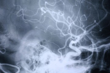
Using specialized CT scans, a team of cardiologists and biomedical engineers developed computer visualizations to see the "shape" of blood flow through the heart's upper left chamber. In a diseased heart, they saw that blood fails to flow in the corkscrew motion that most effectively moves blood out of the left atrium in a healthy heart.
Video credit: Johns Hopkins Medicine
The new study sheds light on just how an enlarged atrium can lead to blood clot formation and increase the risk of stroke. The researchers say the analysis they used could help develop a tool to predict stroke risk in people with heart disease.
A description of the study was published this month in the Annals of Biomedical Engineering.
"By looking at blood flow through the atrium, we think we can accurately assess stroke risk better than such risk factors as heart size and pumping strength," says Hiroshi Ashikaga, assistant professor and member of the Heart and Vascular Institute at Johns Hopkins University's School of Medicine. "Our study fills in a missing diagnostic link between heart function and fluid motion in our understanding of how each can affect stroke risk."
Researchers already knew that enlargement of the heart, particularly the left upper chamber, was linked to increased stroke risk, especially for people with atrial fibrillation, an irregular and often very rapid heart rate. An estimated 1.6 million Americans each year are diagnosed with symptoms of atrial fibrillation that put them at risk for strokes caused by blood clots in the heart.
For their study the Johns Hopkins team compared CT scans of two patients with a history of atrial fibrillation, one with a healthy heart and the other with an enlarged heart. In their computer visualization of the healthy heart, researchers saw that blood flowed in corkscrew motions that circled around into doughnut formations, known as vortexes. The vortexes helped move the blood efficiently through the atrium, with less contact with the atrium's surface tissue.
In the enlarged heart, the researchers saw that at top of the atrium, the blood never fully formed the corkscrews that loop around into vortexes. Instead, the blood seemed to fall in "sheets" that coat the heart's surface.
"The slower the blood moves and the more contact it has with the atrium, the more risk there is for a clot to form," Ashikaga says.
Ashikaga says his team is currently conducting a larger long-term study looking at the blood flow of many more people with normal and ailing hearts, and monitoring the incidence of stroke and other signs of blood clots over time.
Read more from Hopkins MedicinePosted in Health, Science+Technology
Tagged cardiovascular health, heart disease, stroke